 Easy to acquire data for all specimen types
Scanning electron microscopes (SEMs) are indispensable tools not only for research but also for quality assurance and manufacturing sites. At those scenes, the same observation processes need to be performed repeatedly and there has been a need to improve the efficiency of the process.
With the JSM-IT510, the newly added Simple SEM function allows users to "leave the manual repetitive operation to it", required for SEM observation, making SEM observation more efficient and easier.
Features
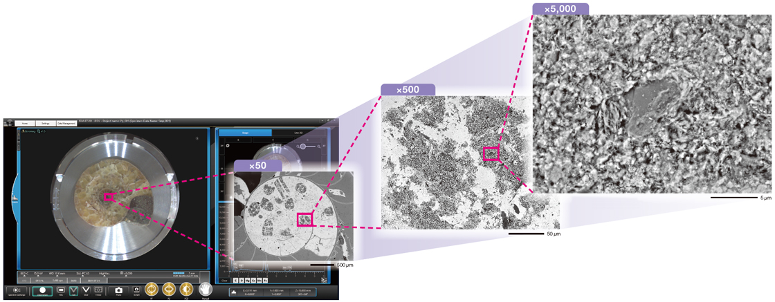
1. Simple SEM
Just select the target field
Simple SEM supports daily routine work.
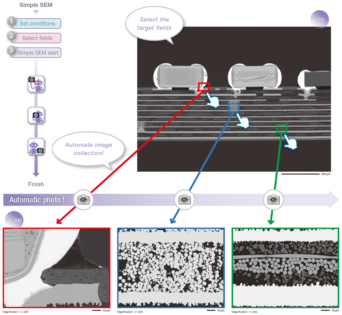
Specimen: Electronic device
Accelerating voltage: 15 kV, (Top) Magnification: ×50 (Bottom) ×1,000, Signal: BE
2. Specimen Exchange Navi
Guide from specimen exchange to automatic observation. Safe and simple!
First, follow the Navi guide to set specimen:
Next, prepare for observation during evacuation:
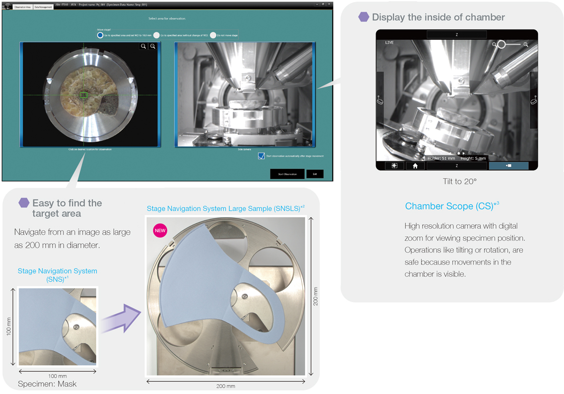
1 Stage Navigation System (SNS) is an option.
2 SNS Large Sample (SNSLS) is an option. Compatible with SNS.
3 Chamber Scope (CS) is an option.
Lastly, start observation automatically, with automatic image formation after evacuation
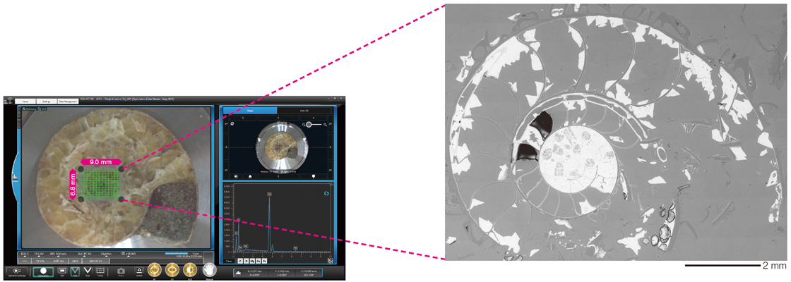
3. Zeromag
Magnify the optical image1, transition to SEM image.
The Zeromag function simplifies navigation providing a seamless transition from the optical to SEM image. The SEM, optical image and holder graphic are all linked for a global view of analysis locations.
1 Stage Navigation System (SNS) is needed to display the optical image.
Specimen: Fossil of ammonite
Accelerating voltage: 7 kV, Signal: BE
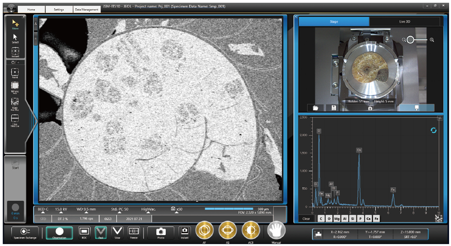
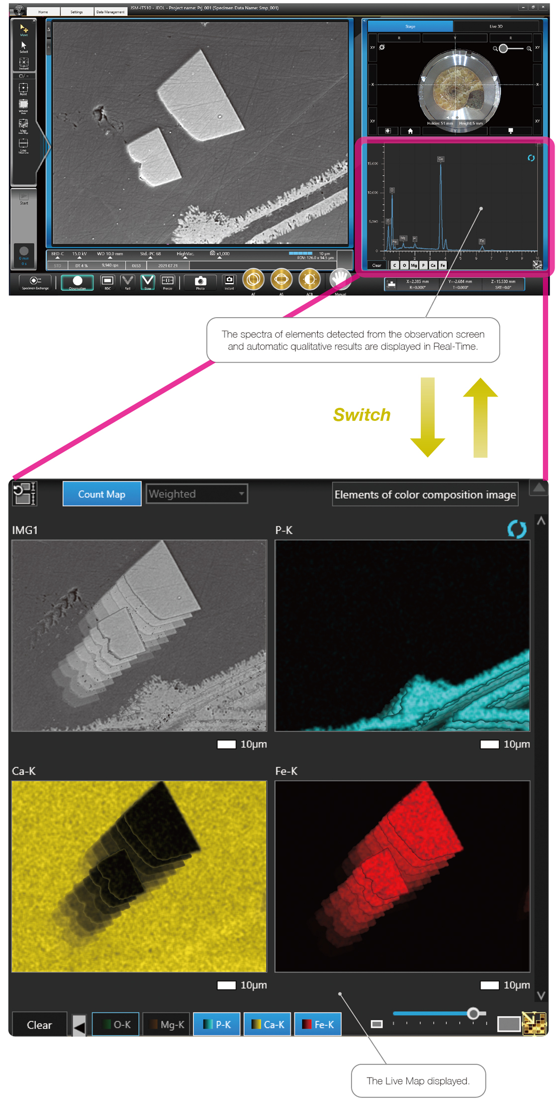
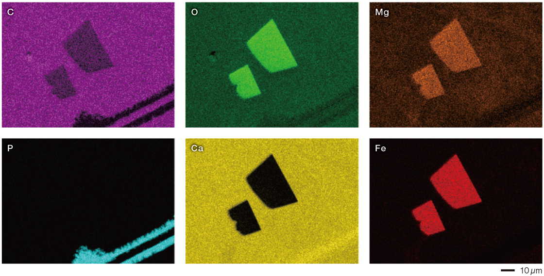
4. Live Analysis / Live Map2
Embedded EDS for Real-Time elemental composition during observation
Live Analysis is a function which displays the EDS spectrum or element maps in Real-Time during image observation. This function can support searching and provide an alert for target elements.
2 Live Analysis is a standard for A (Analysis) / LA (Low Vacuum & Analysis).
Specimen: Fossil of ammonite
Accelerating voltage: 15 kV, Magnification: ×1,000
Simple analysis - the EDS analysis can be started within 3 clicks.
5. Variety of advanced options
Low-vacuum Hybrid Secondary Electron Detector (LHSED)*
This new detector collects both electron and photon signals providing an image with high S/N and enhanced topographic information.
* LHSED is an option. And LV (Low Vacuum) or L A (Low Vacuum & Analysis) is also required.
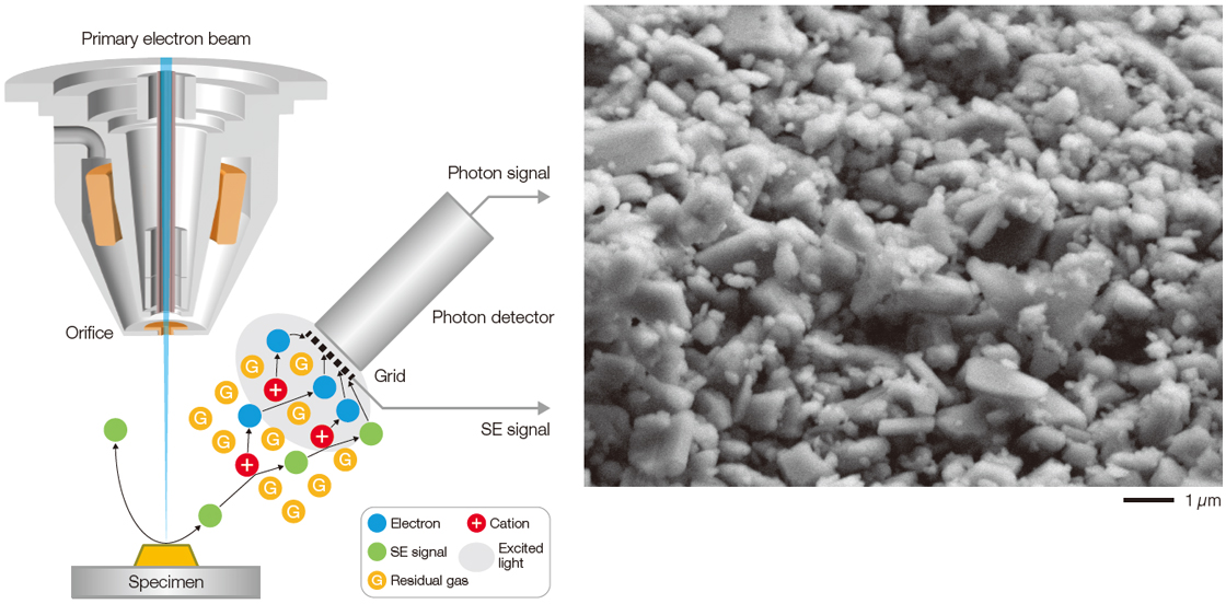
The mechanism of LHSED
Specimen: Plaster
Accelerating voltage: 7 kV, Magnification: x10,000, Signal: LV SE
Live 3D*
The images obtained by a new quadrant BE detector* can be displayed as a live 3D image. 3D images can clearly represent the shape of a specimen, even for those with subtle topographic information.
* Live 3D is a standard in LV (Low Vacuum), LA (Low Vacuum & Analysis). BE detector (option) can be equipped on BU (Base Unit), A (Analysis).
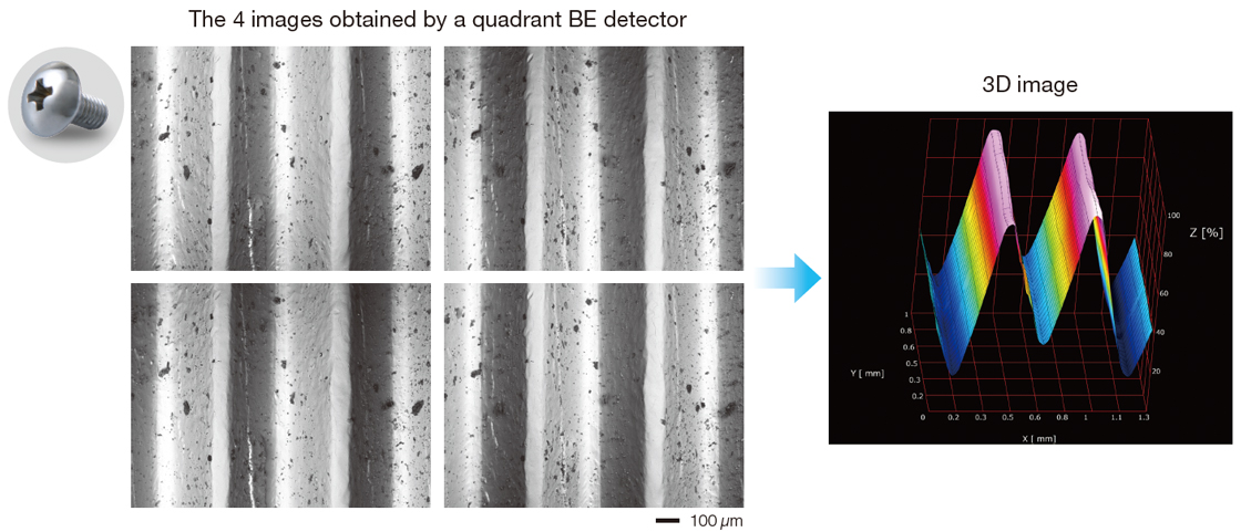
Specimen: Screw
Accelerating voltage: 15 kV Magnification: x100 Signal: BE
Montage
Montage function automates large area image collection and stitching of these images into a composite image.
Specimen: Fossil of ammonite
Accelerating voltage: 15 kV, Magnification: x150, Signal: BE, Number of field: 13 x 13
Stage Navigation System (SNS) is needed to display the optical image.
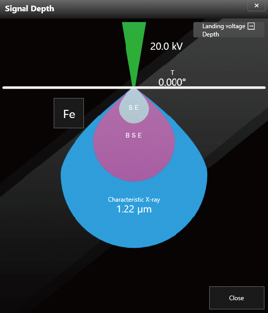
Display the depth of signal
This function displays the analysis depth (approx.) in the specimen. For element analysis, it is very useful.