JMS-T2000GC

JMS-T2000GC

High performance gas chromatograph time-of-flight mass spectrometer.

JEM-ARM300F2

JEM-ARM300F2

A new atomic resolution electron microscope has been released! The "GRAND ARM 2" has been upgraded. This new "GRAND ARM 2" enables observations at ultrahigh spatial resolution with highly sensitive analysis over a wide range of accelerating voltages.

JAM-5200 EBM
CRYO ARM 300 II (JEM-3300)

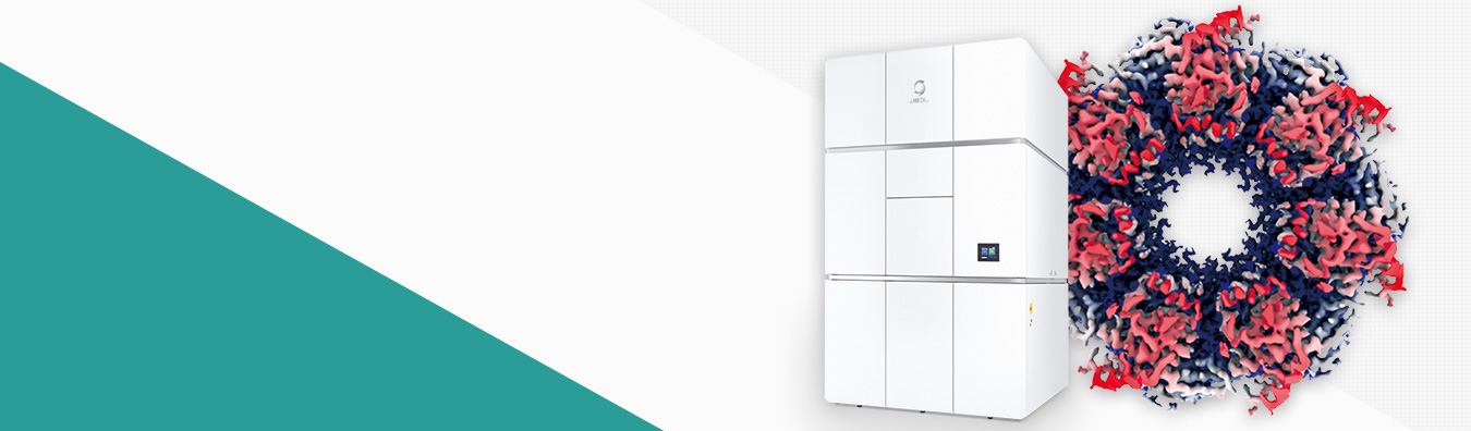
JEM-3300 (CRYO ARM 300 II) Cryo-Electron Microscope

CRYO ARM is an electron microscope for observing biomolecules such as proteins at cryo temperature. The microscope equips a cold field-emission electron gun, an in-column Omega energy filter, a side-entry liquid nitrogen cooling stage, and an automated specimen exchange system. The specimen exchange system can store up to 12 specimens in the specimen exchange chamber.

Electron Microscopy, NMR & Mass Spectometry News

rss

Electron Microscopy News

Our latest blogs on (Scanning) Electron Microscopes, NMR & Mass Spectometry


SuperUser Account
SuperUser Account
SuperUser Account's Blog
Specify Alternate Text

JCM-6000 ”NeoScope™”, A New Desktop Scanning Electron Microscope

JEOL's JCM-5000 NeoScope, since it was announced, has been widely used by electronics manufacturers, including semiconductor device makers, as a desktop scanning electron microscope featuring the same ease of operation as optical microscopes for inspection of the products developed and processed by micro technologies. The new JCM-6000 "NeoScopeTM," is a touch panel controlled, multi functional desktop scanning microscope that answers the increasingly diversified needs among users worldwide. Nikon has joined JEOL to sell the Neoscope.

Features

1) Simple operation

  • Easy touch panel operation
  • A complete range of automated functions (auto focus, auto stigmator, auto contrast/brightness)
  • Easy, dependable auto gun alignment (filament centering)

2) Enhanced low vacuum capability

  • Enhanced quality of backscattered electron images
  • Easy observation of non conductive samples in the direct low vacuum mode
  • Only 2 minutes 30 seconds from sample loading to imaging

3) New capabilities for imaging

  • Secondary electron imaging and backscattered electron imaging supported at high vacuum
  • Dual frame imaging to facilitate comparison of live and retrieved images
  • A wide magnification range from the lowest 10x for wide area of view up to 60,000x

4) A complete line of optional accessories

  • Energy dispersive X-ray spectrometer
  • Tilt/rotation motor drive specimen holder

    *Both options are retrofittable

5) Compact, light, and energy saving

  • Base unit: 330mm (W) x 490mm (D) x 430mm (H); 50 kg
  • Utility: Single phase 100 V to 240 V, 50/60 Hz, 700 to 960 VA
JSM-7100F Thermal field emission electron microscope

JSM-7100F Thermal field emission electron microscope

Observation of nano-structures

The unique high power optics of JSM-7100F guarantees 1.2nm resolution. High magnification for the study of nano-structures is easily obtained.

Stable high precision analysis
The in-lens thermal FEG produces a stable large probe current. You can obtain high quality observation and analysis results. The emitter is guaranteed for 3 years.

Magnetic specimens
The objective lens of the JSM-7100F does not form magnetic field around a specimen. Magnetic specimens can be observed and analyzed without restriction.

Analysis of nano-structures
A small probe diameter is obtained with the patented aperture angle optimizing lens. You can acquire high precision analyses and high quality elemental maps in a short time by using a large probe current.

A variety of analytical equipment including EDS, WDS, and EBSD can be mounted their ideal geometry.

Clean vacuum
A specimen is introduced through the specimen exchange airlock chamber. The specimen chamber is always kept in clean high vacuum. The unique one action specimen exchange mechanism lets you insert and remove a specimen with simple operation. The specimen chamber is pumped with a TMP.

In-lens thermal FEG
The patented in-lens thermal FEG is efficient and produces a maximum probe current of 200nA.

Aperture angle optimizing lens
The patented aperture angle optimizing lens automatically optimizes aperture angle of the objective lens to form a small electron probe diameter at large probe currents.

Observation
A variety of images, such as secondary electron images, backscattered electron images, and STEM images, are obtained. The stable thermal field emission electron source ensures research with high quality images.

Analysis
Elemental analyses with EDS and WDS, and EBSD analysis are carried out efficiently. High precision analysis can be obtained with the stable electron probe.

Specify Alternate Text

Centurio Large Angle SDD-EDS

Centurio is a next-generation Silicon Drift Detector (SDD) EDS that collects X-rays from S/TEM samples at an unprecedented large solid angle of up to 0.98 steradians from a detection area of 100mm2.

Through efficient collection of X-rays at very high count rates, Centurio speeds elemental mapping and improves element detection sensitivity without loss of energy resolution. Large pixel number EDS maps can be made at rates ten times faster than with the previous EDS designs, with excellent signal-to-noise ratio. Combined with the large probe currents in small probe sizes attainable with aberration corrected STEM, fast, efficient atomic resolution EDS analysis is possible.

Key Features

  • High sensitivity, high throughput analysis
  • Exponentially expands the elemental mapping capability for the JEOL 200kV and higher nano-area analysis TEM.
  • The automatically retractable side entry design allows fast repositioning to avoid irradiation from back-scattered electrons.

Specifications

  • Solid angle
    - High resolution polepiece 0.98 sr
    - Ultrahigh resolution polepiece 0.8 sr
  • Detection area 100mm2
Specify Alternate Text

JEOL Announces a New Field Emission Scanning Electron Microscope, JSM-7800F

An ultimate analytical tool for a wide range of applications

Scanning electron microscopes are used in a wide range of fields from basic material research to specific applications in production. The JSM-7800F is an ultimate analytical tool that answers a wide variety of users' needs. With its new super hybrid objective lens, the microscope is capable of high resolution imaging as well as high speed, high precision elemental analysis.

Features

Ultimate resolution

The super hybrid objective lens achieves resolutions of 0.8 nm (15 kV) and 1.2 nm (1 kV).

High speed, high precision elemental analysis

The microscope performs speedy sample analysis with the optimally focused electron probe at high current without compromising the analytical accuracy and the quality of elemental mapping.

High quality data acquired by stable electron probe

The long lasting in-lens thermal electron gun produces a stable probe, which allows for continuous acquisition of high quality data from imaging and from various analysis including EDS, WDS, EBSD, and CL.

Support of wide ranging samples

The super hybrid objective lens can image/analyze magnetic samples at high magnification. It also makes imaging of non conductive samples easy.

Specify Alternate Text

Multi-touch Interface Creates New Electron Microscope Experience with JEOL InTouch Scope™

Logo InTouch ScopeJEOL offers a whole new electron microscope experience with the introduction of the InTouch Scope™, an analytical, low vacuum Scanning Electron Microscope (SEM) featuring integrated Energy Dispersive Spectroscopy (EDS) with the latest Silicon Drift Detector (SDD) technology.

The new InTouch Scope has the familiar feel of today’s personal electronic media. The intuitive multi-touch screen interface puts all SEM “Apps” at the operator’s fingertips. The user can expand windows and images with the sweep of two fingers, dial in magnification and focus with a swipe, and select operating parameters, analytical functions, or measure distances just by tapping the PC or notebook touch screen.

Ease of use is a key feature of all JEOL SEMs, and the versatile InTouch Scope has functions that users of all levels will appreciate:

  • automatic SEM condition setup based on sample type
  • simultaneous multiple live image and movie capture
  • easy sample navigation at 5x – 300,000x magnifications
  • quantitative and qualitative elemental analysis
  • low and high vacuum operation
  • Wireless capability.

The In-Touch Scope features all the capabilities of a full size tungsten SEM with integrated EDS analysis in a small, ergonomic and intuitive design. An onboard turbo pump make this a truly self-contained, portable SEM that is easy to set up anywhere in the lab.

Specify Alternate Text

JEOL Reinvents Time-of-Flight Mass Spectrometry with Innovative SpiralTOF™ MALDI-TOF System

JEOL announced the introduction of the company’s first commercially available MALDI-TOF mass spectrometer, the JEOL JMS-S3000 SpiralTOF™. The SpiralTOF reinvents Time-of-Flight ion optics with an extended flight length in a compact footprint, delivering a resolving power of greater than 60,000 (FWHM) over a wide mass range of m/z 10-30,000 - the highest resolving power of all commercial MALDI-TOF systems.

JEOL’s patented technology consists of a staggered figure-8 ion trajectory of 17 meters. By refocusing the ion packets during each turn, the divergence of the ions is reduced over the flight distance. The SpiralTOF ion optical system is engineered to overcome the limits of delayed extraction with kinetic energy convergence resulting in high sensitivity, resolving power and mass accuracy (less than 1ppm) over a wide mass range.

To meet the diverse needs of researchers, the SpiralTOF is available in four configurations:

  • MALDI-TOFMS with unsurpassed high mass resolving power and high mass accuracy
  • Linear TOF option for high sensitivity analysis of high molecular weight samples and/or those that undergo post-source decay
  • TOF/TOF option for acquiring high energy collision-induced dissociation (CID) product ion spectra of monoisotopically selected precursor ions
  • TOF/TOF plus Linear TOF for full analytical capability

With the addition of the SpiralTOF to its mass spectrometer product line, JEOL makes available a unique analytical instrument for proteomics, synthetic polymers, materials, and biomolecules.

JEOL offers a full suite of mass spectrometry systems: the first commercially-available open air ion source, Direct Analysis in Real Time (DART®), paired with its AccuTOFTM-DART Time-of-Flight mass spectrometer; the GC-TOF AccuTOF-GCv mass spectrometer; the JEOL MStation double focusing magnetic sector mass spectrometer; and the GCMate II benchtop GC/MS. Additionally, a family of 300 to 930MHz NMR spectrometers completes its chemical analysis product line.

Specify Alternate Text

First JEOL JEM-ARM200F Electron Microscope Produces Atomic Resolution Data in Record Time at University of San Antonio

The first transmission electron microscope of its caliber to be installed, the eagerly awaited atomic resolution JEOL JEM-ARM200F TEM arrived at the University of Texas at San Antonio in January, and by early February began producing outstanding imaging results.
“Achieving raw HAADF images showing at least 78 picometer information transfer in just three weeks demonstrates the stability of this all-new instrument and the skill of the UTSA-JEOL team to quickly power up the first TEM of its kind,” said Dr. Thomas Isabell, JEOL USA Director of the TEM Product Division.

UTSA physics and astronomy department chair Dr. Miguel Yacaman, a renowned electron microscopist and nanotechnology researcher, tested the new ARM200F performance on Si <110> samples as the JEOL engineering, service and applications team worked closely together to install the TEM in UTSA’s Advanced Microscopy Lab.

“At the level of this new microscope, the potential for new discoveries is enormous,” says Yacaman, who likened the capabilities of the JEOL ARM200F for sub-atomic research to those of the Hubble telescope for intergalactic exploration.

JEOL ARM200F Electron Microscope - Atomic Resolution Data

The ARM200F represents more than 60 years of TEM expertise at JEOL and was designed from the ground up to integrate aberration correction into a super-shielded electron column that safeguards the ultrahigh-powered optics from environmental interferences.

“This first installation of the ARM200F demonstrates that JEOL is out in front in the development of stable, ultra-quality instruments and showcases just what our service and applications teams are capable of.” said Isabell.

The JEM-ARM200F enables both atom-by-atom imaging resolution and unmatched spatial resolution for atom-to-atom chemical mapping of materials, including EDS (energy-dispersive x-ray spectroscopy) and EELS (electron energy-loss spectroscopy). The completely new electron column design integrates S/TEM with Cs correction for atomic spatial energy resolution combined with high probe currents for microanalysis.

Specify Alternate Text

ClairScope™ (JASM-6200)

JEOL Ltd. (Gon’emon Kurihara, President) announced a new hybrid microscope, JASM-6200 “ClairScope™” to be distributed in August 2009.

Design Principle

ClairScope™ consists of an atmospheric scanning electron microscope (ASEM) and an optical microscope positioned on top. The ASEM is isolated from the SEM with a thin film installed at the top end of the inverted SEM. The thin film, designed to transmit an electron beam while blocking air, separates the sample in atmosphere from the vacuum in the SEM. The electron beam is projected from below to the sample placed on the thin film for high resolution imaging of the sample in atmosphere. The same area of view of the sample can also be imaged in the optical microscope on top. The thin film is configured in a dish (thin film dish), which can be used for cell culture in a culture chamber.

Features

SEM imaging in complete atmospheric pressure

The sample is held in complete atmospheric pressure, enabling dynamic observation of physical and chemical reactions in liquids and gases. Lengthy preliminary treatment of biological samples, including dehydration and drying, is no longer necessary, resulting in high throughput imaging. The microscope, without the limitations of vacuum atmosphere, will broaden the range of applications.

Imaging of the same area of view as optical microscope

The top of the thin film is open, joined to the optical microscope. The optical microscope being aligned with the ASEM, the operator can image the same area of view alternately between both microscopes.

The optical microscope can accommodate up to 6 mirror units simultaneously. Standard mirror units for bright field imaging and ultraviolet irradiation, combined with optional mirror units, can acquire various types of fluorescent images.

Open specimen chamber

The specimen chamber is open, allowing the operator to externally control reagents (chemical administration).For example, the operator can load the thin film dish onto the system, and observe the sample in the ASEM after administering a chemical to the sample. The operator can also monitor physical and chemical developments as the sample changes its volume.

Biological applications

Extended applications of optical microscope

Many biologists routinely use optical microscopes for their research. However, optical microscopy is unable to achieve resolution higher than 0.2 um due to its wavelength limitations. While scanning electron microscopy (SEM) and transmission electron microscopy (TEM) feature higher resolution, many samples require a lengthy preliminary treatment process including multiple dehydration and drying process steps, which usually takes a skilled technician one to a few days to complete. This is why SEM and TEM, despite their high resolution, are not as widely used as optical microscopy.

ClairScope™ is designed to observe samples in atmospheric pressure using the ASEM. It is capable of high resolution imaging of biological samples without the preliminary dehydration/drying process that requires skilled technician. Sample pre-treatment consists of simple steps of chemical administration, taking only about 10 minutes. This enhances the efficiency and yield of sample imaging.

ClairScope™ also supports imaging of the same area of view in the optical microscope on top and the ASEM at the bottom. This allows optical microscope users to first observe images they are familiar with, and proceed to high resolution imaging of a given spot of the sample. The system allows the operator to identify tissues and local existence of protein using fluorescence staining and optical microscopy, and to further study the corresponding spots using high resolution imaging of the ASEM.

ClairScope™ also allows the operator to observe live cells in the optical microscope, restrict the motion with chemicals, and apply fixation/staining as needed for high resolution imaging in the ASEM. Cells are cultured on the thin film dish in the same way as any conventional plastic dish. The researcher can culture cells in the usual procedure, observe the cells in the optical microscope, and focus on areas of interests in the ASEM for further imaging.

ClairScope™ is expected to be widely used in a variety of fields including basic biology, medicine, pharmaceutical, and cosmetics.

Other applications

Dynamic observation of chemical reaction

Real time high resolution imaging can reveal the mechanism of chemical reactions in liquid and vapor. Traditionally, this has been accomplished by TEM with an environmental cell. However, TEM can only accept thin film samples, and SEM imaging has been in demand for certain samples. ClairScope™, designed to image samples in atmospheric pressure in the ASEM, is capable of real time imaging of chemical reactions in liquid and vapor. The open top of the thin film facilitates chemical administration, and enables observation of gas emitting reactions. Furthermore, the system allows the optical microscope on top to alternately observe the same area of view as the ASEM. The system has succeeded in dynamic observation of hydrolytic reactions of plaster.

Dynamic observation of drying process

Drying is a critical process in various industries. However, it has been difficult to achieve dynamic observation of the drying process at higher resolution than optical microscopy. ClairScope™, with the open top of the thin dish, enable observation of the drying process of solvents. It has succeeded in dynamic observation of the drying process of a salt containing solvent from drying to crystallization.

Dynamic observation of electrochemical reaction

Electrochemical reactions, related to batteries, plating, corrosion, and refinement, is essential in many industries. High resolution imaging of electrochemical reactions in liquid has been a difficult issue. ClairScope™, capable of real time high resolution imaging of reactions in liquid in atmospheric pressure in the ASEM, also supports dynamic observation of electrochemical reactions. With the open top of the dish, the system is also effective in observing electrochemical reactions generating gases. It has succeeded in dynamic observation of electrochemical reactions of a sample loaded onto a prototype thin film dish incorporating an electrode in the thin film.

Basic physics

Particles in liquid are known to demonstrate Brownian motion and self assembly under certain conditions. These reactions are related to the basic motion mechanism of life, a field to be explored extensively. These reactions have been studied in optical microscopy. ClairScope™ has succeeded in real time observation of Brownian motion and self assembly of particles.

Reaction to electron beam irradiation

A high intensity electron beam can induce reactions in liquid and vapor. Using ClairScope™, the operator can induce such reaction in the ASEM for dynamic imaging, and also monitor the reaction using the optical microscope on top.

Specify Alternate Text

NeoScope Benchtop SEM voted European Best New Lab Product

The NeoScope benchtop SEM, born from the combined expertise of Nikon Instruments and JEOL, is the winner of the SelectScience Scientists’ Choice Award for European Best New General Lab Product 2008.

Announced at the recent ACHEMA exhibition in Germany, the Scientists' Choice Awards were based on the results of an independent poll of more than 25,000 scientists from the SelectScience online community, who were asked to nominate and vote for what they considered to be the best new products of 2008. SelectScience.net is the leading online resource for applied chemists, clinical chemists and life scientists.

“Scientists are always quick to embrace innovation and new technology,” Chay Keogh, Marketing Manager at Nikon instruments, “The NeoScope is a fresh new approach offering a powerful yet affordable benchtop SEM. The Scientist’s Choice Award confirms that our combined experience of over 150 years in pioneering imaging technology has enabled JEOL and Nikon to lead the way in providing another exciting new tool to help accelerate the pace of research in all fields.”

The SelectScience Choice Award adds to Nikon’s exclusive Perfect Focus System second place ranking in The Scientist’s top innovations of 2008.

View the Select Science Awards presentation at ACHEMA

Specify Alternate Text

JEM-ARM200F, An Ultimate Atomic Resolution Transmission Electron Microscope

JEOL Ltd. (Gon’emon Kurihara, President) announced a new transmission electron microscope, JEM-ARM200F, incorporating a spherical aberration corrector for the electron optic system as standard, to be distributed in March 2009.

Background

Transmission electron microscopes are designed to study the geometry and structure of substances at high resolution. They can also analyze the elements constituting substances and the status of electrons by incorporating various analyzers such as energy dispersive X-ray fluorescent spectrometer (EDS) and electron beam energy loss spectrometer (EELS).

A new technology has recently been introduced to correct spherical aberrations that have long restricted the resolving power of electron microscopes. Spherical aberration correctors significantly enhance the resolution and analytical capabilities, enabling ultimate atomic level analysis. The JEM-ARM200F is a powerful transmission electron microscope with a standard spherical aberration corrector, featuring sub angstrom resolution and atomic level analysis.

Features

  • Atomic level resolution - STEM: 0.08 nm; TEM: 0.11 nm
  • Standard spherical aberration corrector for electron optic system eliminating spherical aberrations
  • Maximum accelerating voltage 200 kV